Beranda
/ Loculated Pleural Effusion Meaning / Pleural Effusion Parapneumonic Md Nexus : Loculated pleural effusion is probably the most common cause of this appearance.
Loculated Pleural Effusion Meaning / Pleural Effusion Parapneumonic Md Nexus : Loculated pleural effusion is probably the most common cause of this appearance.
Insurance Gas/Electricity Loans Mortgage Attorney Lawyer Donate Conference Call Degree Credit Treatment Software Classes Recovery Trading Rehab Hosting Transfer Cord Blood Claim compensation mesothelioma mesothelioma attorney Houston car accident lawyer moreno valley can you sue a doctor for wrong diagnosis doctorate in security top online doctoral programs in business educational leadership doctoral programs online car accident doctor atlanta car accident doctor atlanta accident attorney rancho Cucamonga truck accident attorney san Antonio ONLINE BUSINESS DEGREE PROGRAMS ACCREDITED online accredited psychology degree masters degree in human resources online public administration masters degree online bitcoin merchant account bitcoin merchant services compare car insurance auto insurance troy mi seo explanation digital marketing degree floridaseo company fitness showrooms stamfordct how to work more efficiently seowordpress tips meaning of seo what is an seo what does an seo do what seo stands for best seotips google seo advice seo steps, The secure cloud-based platform for smart service delivery. Safelink is used by legal, professional and financial services to protect sensitive information, accelerate business processes and increase productivity. Use Safelink to collaborate securely with clients, colleagues and external parties. Safelink has a menu of workspace types with advanced features for dispute resolution, running deals and customised client portal creation. All data is encrypted (at rest and in transit and you retain your own encryption keys. Our titan security framework ensures your data is secure and you even have the option to choose your own data location from Channel Islands, London (UK), Dublin (EU), Australia.
Loculated Pleural Effusion Meaning / Pleural Effusion Parapneumonic Md Nexus : Loculated pleural effusion is probably the most common cause of this appearance.. The lack of specificity is mainly due to the limitations of the imaging modality. Often it happens in the context of a pneumonia, injury, or chest surgery. Fibrinopurulent, when fibrous septa form localized. Loculated malignant effusions however, are inherently resistant to the usual approaches because of nonexpanding underlying lung. Causes of an exudative effusion are malignancy, infection, or inflammatory disorders such as rheumatoid arthritis.
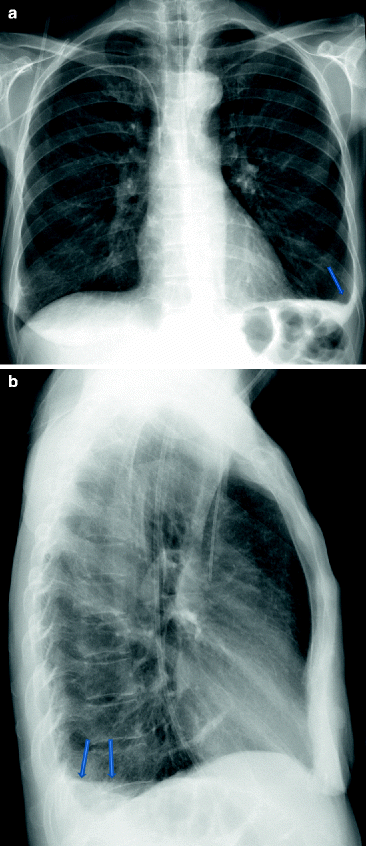
The space where the fluid is located is called the pleura, and it plays a vital role in the health and function of the lungs as well as the rest of the respiratory system. Obliteration of left costophrenic angle with a wide pleural based dome shaped opacity projecting into the lung noted tracking along the cp angle and lateral chest wall suggestive of loculated pleural effusion, however the possibility of empyema can not be ruled out completely. Often it happens in the context of a pneumonia, injury, or chest surgery. The pleura is a thin piece of tissue with 2 layers. Divided into small spaces, compartments or cavities.
Right Subpulmonic Pleural Effusion Images Diagnosis Treatment Options Answer Review Thoracic Imaging from vcuthoracicimaging.com If your doctor suspects a malignant pleural effusion, the next step is usually a thoracentesis, a procedure in which a needle is inserted through the chest wall into the pleural space to get a sample of the fluid. Most malignant effusions can be controlled by thoracentesis and/or closed thoracostomy tube drainage and sclerosis of the pleural cavity. Treatment may fail if the catheter is not placed optimally within the loculation or if the fluid is hemorrhagic or fibrinous. Obliteration of left costophrenic angle with a wide pleural based dome shaped opacity projecting into the lung noted tracking along the cp angle and lateral chest wall suggestive of loculated pleural effusion, however the possibility of empyema can not be ruled out completely. But clinical identification of pleural effusion is possible only when the amount of fluid is more than 500ml. Most effusions start like this and can be easily missed. Fibrinopurulent, when fibrous septa form localized. In chf effusions are bilateral and more on right.
But clinical identification of pleural effusion is possible only when the amount of fluid is more than 500ml.
Loculated pleural effusion is probably the most common cause of this appearance. A pleural effusion is the accumulation of excess fluid in the pleural cavity surrounding the lungs. The lack of specificity is mainly due to the limitations of the imaging modality. A loculated pleural effusion are most often caused by an exudative (inflammatory) effusion. (1)department of thoracic medicine, university hospital, heraklion, crete, greece. The space where the fluid is located is called the pleura, and it plays a vital role in the health and function of the lungs as well as the rest of the respiratory system. Use of pleural ph assumes that a single measurement conveys a representative picture of ph throughout the effusion. Fibrinopurulent, when fibrous septa form localized. Transudate is usually composed of ultrafiltrates of plasma due to an imbalance in vascular. But clinical identification of pleural effusion is possible only when the amount of fluid is more than 500ml. Surgical thoracostomy tube placement and radiologically guided catheter drainage are standard therapy for loculated pleural fluid collections. Loculated malignant effusions however, are inherently resistant to the usual approaches because of nonexpanding underlying lung. Exudative, when there is an increase in pleural fluid with or without the presence of pus;
The space where the fluid is located is called the pleura, and it plays a vital role in the health and function of the lungs as well as the rest of the respiratory system. This type of effusion is empyema unless proven otherwise. Most malignant effusions can be controlled by thoracentesis and/or closed thoracostomy tube drainage and sclerosis of the pleural cavity. A pleural effusion is a buildup of fluid between the layers of tissue that line the lungs and chest cavity. (1)department of thoracic medicine, university hospital, heraklion, crete, greece.
Pleural Effusion Pyothorax Pneumothorax Ppt Video Online Download from slideplayer.com A 2 effusions (those with a poor prognosis) occupy more than 50% of the hemithorax, are loculated, and/or are associated with thickening of the parietal pleural. Surgical thoracostomy tube placement and radiologically guided catheter drainage are standard therapy for loculated pleural fluid collections. The other rests on the chest wall. What are the different appearances of pleural effusion? The space where the fluid is located is called the pleura, and it plays a vital role in the health and function of the lungs as well as the rest of the respiratory system. A systematic approach to analysis of the fluid in. Decortication may be performed using small incisions (thoracoscopy) or a large one (thoracotomy). The pleura are thin membranes that line the lungs and the inside of the chest cavity and act to lubricate and facilitate breathing.
Most effusions start like this and can be easily missed.
Pleural empyema is a collection of pus in the pleural cavity caused by microorganisms, usually bacteria. The other rests on the chest wall. Divided into small spaces, compartments or cavities. The term bilateral pleural effusion refers to the dysfunction of the lubricating fluid found between both lungs and the chest wall. The first symptoms of pleural effusion are usually seen when the amount of fluid in the pleura reaches about 500 milliliters. The space where the fluid is located is called the pleura, and it plays a vital role in the health and function of the lungs as well as the rest of the respiratory system. Use of pleural ph assumes that a single measurement conveys a representative picture of ph throughout the effusion. A pleural effusion is a collection of fluid in the space between your chest wall and lungs. Role of streptokinase in the treatment of acute loculated parapneumonic pleural effusions and empyema. If your doctor suspects a malignant pleural effusion, the next step is usually a thoracentesis, a procedure in which a needle is inserted through the chest wall into the pleural space to get a sample of the fluid. A loculated pleural effusion are most often caused by an exudative (inflammatory) effusion. A pleural effusion is accumulation of excessive fluid in the pleural space, the potential space that surrounds each lung. A systematic approach to analysis of the fluid in.
Pleural effusion is fluid buildup in the space between the layers of the pleura. The first symptoms of pleural effusion are usually seen when the amount of fluid in the pleura reaches about 500 milliliters. (1)department of thoracic medicine, university hospital, heraklion, crete, greece. Icu patients cannot sit up and the effusion layers posteriorly. The other rests on the chest wall.
Abnormalities Involving The Pleura Springerlink from media.springernature.com Pleural effusions in the intensive care setting. Pleural effusion is fluid buildup in the space between the layers of the pleura. 3.7, a ), hemorrhage, and neoplasms. Often effusions are multiloculated, and varying concentrations of nondiffusible acids such as lactic acid, if present in different concentrations, could mean clinically significant variations in ph between locules. An excessive accumulation of fluid in the pleural space is known as a pleural effusion. Loculated malignant effusions however, are inherently resistant to the usual approaches because of nonexpanding underlying lung. The type of fluid that forms a pleural effusion may be categorized as either transudate or exudate. Bouros d(1), schiza s, panagou p, drositis j, siafakas n.
We studied the value of transca …
A pleural effusion is a collection of fluid in the space between your chest wall and lungs. A pleural effusion is a buildup of fluid in the pleural space, an area between the layers of tissue that line the lungs and the chest wall. Surgical thoracostomy tube placement and radiologically guided catheter drainage are standard therapy for loculated pleural fluid collections. (1)department of thoracic medicine, university hospital, heraklion, crete, greece. Divided into small spaces, compartments or cavities. The space where the fluid is located is called the pleura, and it plays a vital role in the health and function of the lungs as well as the rest of the respiratory system. Bouros d(1), schiza s, panagou p, drositis j, siafakas n. Loculated effusions, defined as effusions that do not shift freely in the pleural space, occur when there are adhesions between the visceral and parietal pleura. The other rests on the chest wall. The pleura is a thin piece of tissue with 2 layers. Icu patients cannot sit up and the effusion layers posteriorly. Often it happens in the context of a pneumonia, injury, or chest surgery. A pleural effusion is a buildup of fluid between the layers of tissue that line the lungs and chest cavity.
Pulmonology 16 years experience see below: loculated pleural effusion. Transudate is usually composed of ultrafiltrates of plasma due to an imbalance in vascular.